Diagram Of Shoulder Joint / - The shoulder joint (glenohumeral joint) is a ball and socket joint between the scapula and the humerus.
byElnora Webb•
0
Diagram Of Shoulder Joint / - The shoulder joint (glenohumeral joint) is a ball and socket joint between the scapula and the humerus.. Atlas of the anatomy of the joint of the shoulder on a ct arthrogram in axial, coronal, and sagittal sections, on a 3d images and on conventional athrogram. Where the rounded top of the arm bone (humerus) contacts the shoulder blade is called the glenohumeral joint. This diagram here just shows the joint capsule itself. Nerve innervation of the shoulder joint. The pairing of shoulder internal rotation and protraction may be common in postural dysfunction, but.
Dislocation of the shoulder is extremely painful and may require surgical repair or even cause permanent damage. The shoulder joint is supplied with blood by branches of the anterior and posterior circumflex humeral arteries, the suprascapular artery and the scapular circumflex diagram of the human shoulder joint, back view. The shoulder joint is vulnerable to dislocations from sudden jerks of the arm, especially in children before strong muscles have developed. Editor · aug 8, 2017 ·. We can also call this adduction of the scapulae. protraction is the pulling forward of the shoulder joint.
Soft Tissues of the Shoulder from embed.widencdn.net Diagram of the human shoulder joint. It can also be called abduction as the movement pulls the scapula away from the vertebrae. Just remember the articulating surfaces. It is formed by the head of the humerus and the glenoid cavity of the scapula. This is called the glenoid. Here, we shall consider the factors the permit movement, and. Check out this shoulder joint science diagram template in the edraw free download resources library. Muscle diagram of shoulder shoulder muscles diagram shoulder muscle diagrams diagram site.
The shoulder joint is vulnerable to dislocations from sudden jerks of the arm, especially in children before strong muscles have developed.
Comprising of numerous ligamentous and muscular structures, the only actual bony articulations are the glenohumeral joint and the acromioclavicular jo. The shoulder joint may be subjected to different injuries. Here, we shall consider the factors the permit movement, and. This diagram here just shows the joint capsule itself. Deep muscles of shoulder at temple university,shoulder joint anatomy pictures and information,dr. Russell janssen, chiropractor in clearwater of tampa bay. It is a ball and socket joint that allows the arm to rotate in a circular fashion or to hinge out. The first type is the white cartilage on the ends of the bones (called articular cartilage) which allows the bones to glide and move on each other. Posted on november 17, 2018november 17, 2018. Retraction pulls the shoulder joint to the rear and toward the vertebral column. Learn vocabulary, terms and more with flashcards, games and other study tools. Click now and learn everything about its anatomy and function at kenhub! You can see it enclosing the glenohumeral joint and you can see its attachment on the anatomical neck that's the shoulder joint.
The glenohumeral joint is the main joint of the shoulder and the generic term shoulder joint usually refers to it. The shoulder joint may be subjected to different injuries. The shoulder joint is vulnerable to dislocations from sudden jerks of the arm, especially in children before strong muscles have developed. Human shoulder joint pain anatomy. The glenohumeral, or shoulder, joint is a synovial joint that attaches the upper limb to the axial skeleton.
Shoulder diagram | Healthiack from healthiack.com Shoulder joint is the most mobile joint of the human body. This page is about diagram muscle shoulder joint,contains upper extremity occupational therapy 205 with teresa at tufts university,2. Simply put, the shoulder, or shoulder joint, is the connection of the upper arm and the thorax. The shoulder joint is vulnerable to dislocations from sudden jerks of the arm, especially in children before strong muscles have developed. Shoulder joint anatomy and information the most flexible joint in the entire human body our shoulder joint is formed by the union of the humerus the scapula or shoulder blade and the shoulder muscles anatomy diagram & function. Clinical examples of shoulder joint injuries or lesions. Know every tiny but important part of your arms from the humeral head to the scapula. The glenohumeral joint is the main joint of the shoulder and the generic term shoulder joint usually refers to it.
It is formed by the head of the humerus and the glenoid cavity of the scapula.
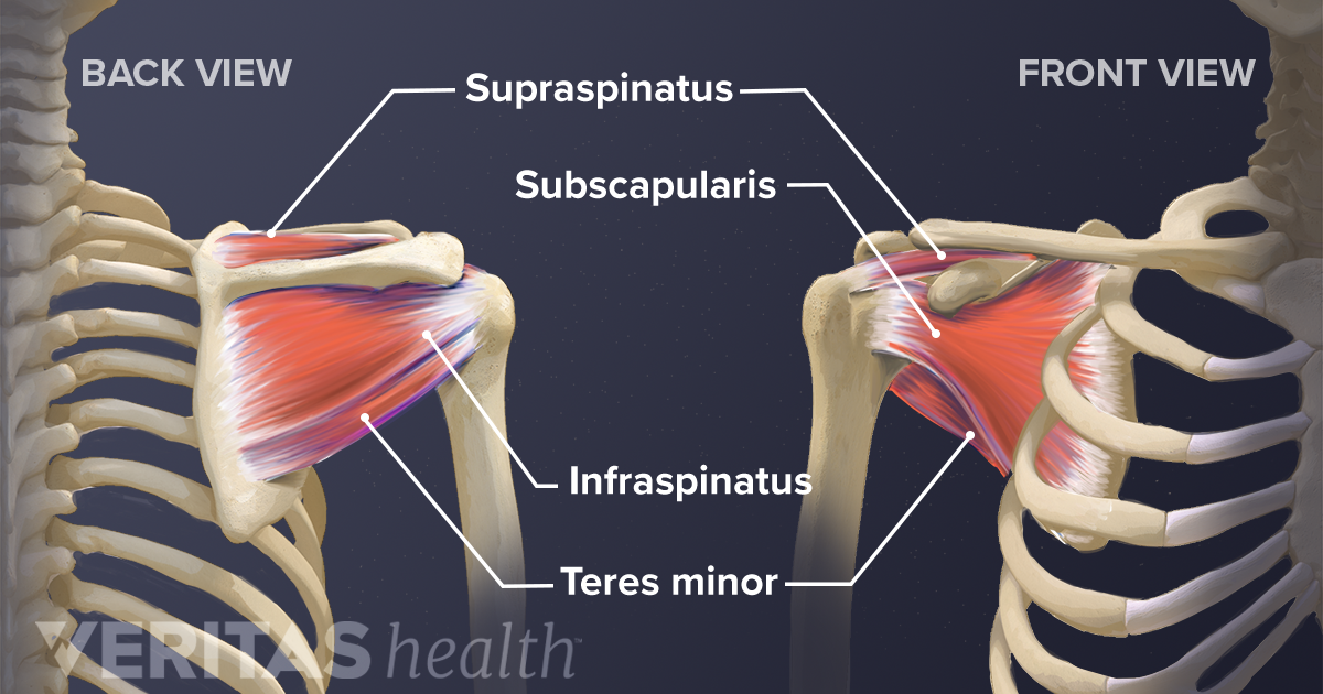
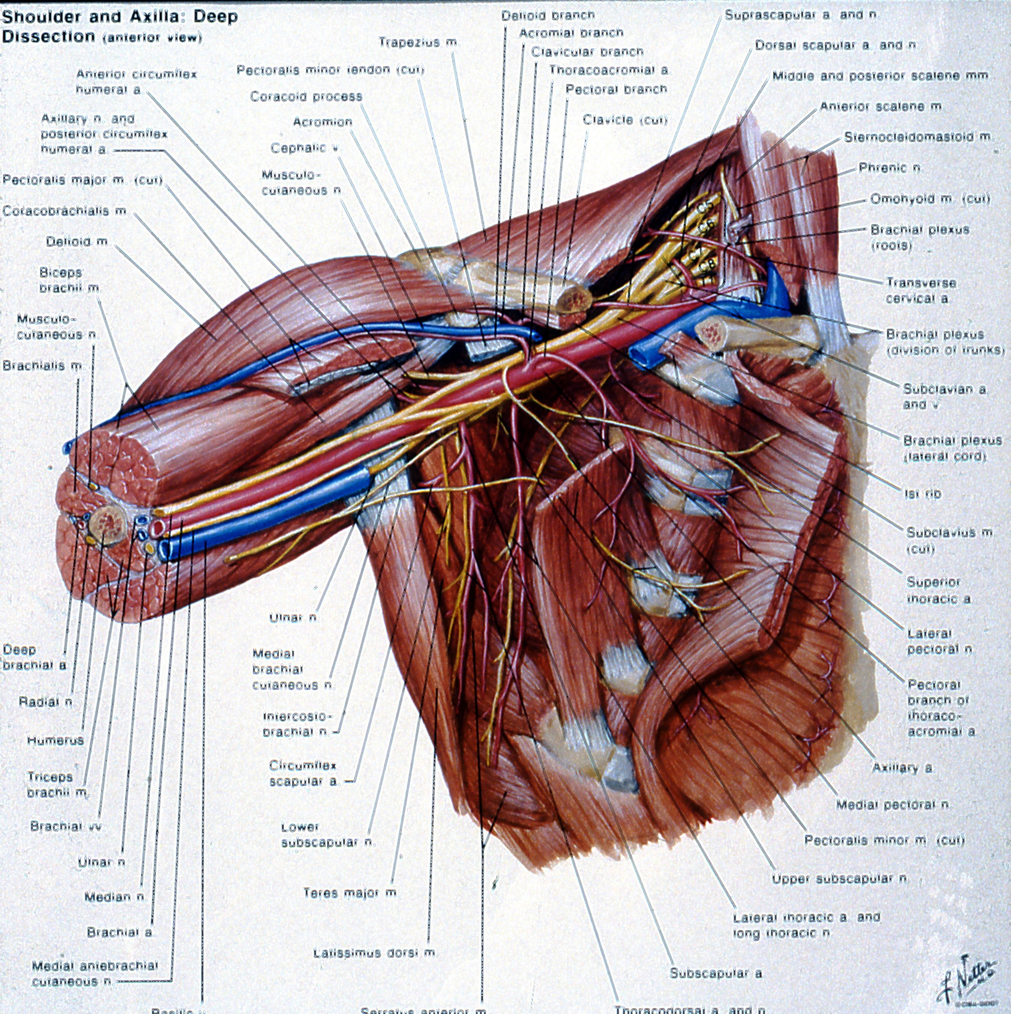
It is formed by the head of the humerus and the glenoid cavity of the scapula. The supraspinatus, infraspinatus, teres minor, and subscapularis muscles and their tendons comprise the rotator cuff, and. Check out this shoulder joint science diagram template in the edraw free download resources library. Editor · aug 8, 2017 ·. Deep muscles of shoulder at temple university,shoulder joint anatomy pictures and information,dr. It is a ball and socket joint that allows the arm to rotate in a circular fashion or to hinge out. Suprascapular , axillary, subscapular , lateral pectoral and musculocutaneous nerve branches. This diagram here just shows the joint capsule itself. Shoulder joint is the most mobile joint of the human body. Clinical examples of shoulder joint injuries or lesions. Retraction pulls the shoulder joint to the rear and toward the vertebral column. We can also call this adduction of the scapulae. protraction is the pulling forward of the shoulder joint. Simply put, the shoulder, or shoulder joint, is the connection of the upper arm and the thorax.
Here, we shall consider the factors the permit movement, and. It involves articulation between the glenoid cavity of the scapula (shoulder blade). Know every tiny but important part of your arms from the humeral head to the scapula. There are two kinds of cartilage in the joint. Shoulder joint is the most mobile joint of the human body.
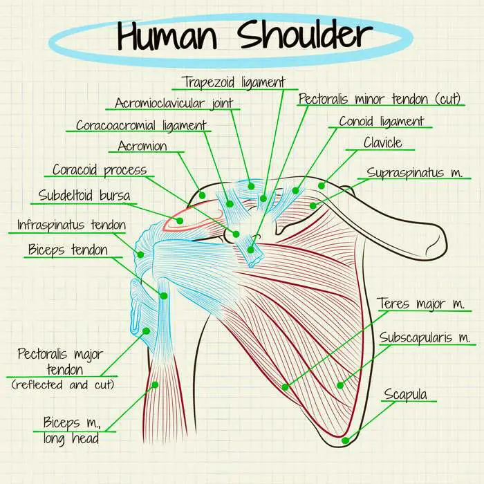
Stuart Kozinn, MD - Scottsdale Joint Center from scottsdalejointcenter.com Following inferior dislocation of shoulder joint, the rounded contour of shoulder is lost and there is weakness of abduction of armbecause the. It is a ball and socket joint that allows the arm to rotate in a circular fashion or to hinge out. Shoulder joint anatomy and information the most flexible joint in the entire human body our shoulder joint is formed by the union of the humerus the scapula or shoulder blade and the shoulder muscles anatomy diagram & function. Shoulder is composed of 4 joints, namely glenohumeral or shoulder joint, acromioclavicular joint, sternoclavicular joint and scapulothoracic joint. The background music used in the. Just remember the articulating surfaces. We can also call this adduction of the scapulae. protraction is the pulling forward of the shoulder joint. Shoulder joint of human body anatomy infographic diagram with all parts including bones ligaments muscles bursa cavity capsule cartilage membrane for medical science education and health care.
Following inferior dislocation of shoulder joint, the rounded contour of shoulder is lost and there is weakness of abduction of armbecause the.
Shoulder joint of human body anatomy infographic diagram with all parts including bones ligaments muscles bursa cavity capsule cartilage membrane for medical science education and health care. The shoulder joint is supplied with blood by branches of the anterior and posterior circumflex humeral arteries, the suprascapular artery and the scapular circumflex diagram of the human shoulder joint, back view. Dislocation of the shoulder is extremely painful and may require surgical repair or even cause permanent damage. A selection of frequent lesions encountered by medical students as well as skilled orthopedists and surgeons, and neurologists, is presented here. Clinical examples of shoulder joint injuries or lesions. Retraction pulls the shoulder joint to the rear and toward the vertebral column. Russell janssen, chiropractor in clearwater of tampa bay. Posted on november 17, 2018november 17, 2018. The glenohumeral joint is the main joint of the shoulder and the generic term shoulder joint usually refers to it. Simply put, the shoulder, or shoulder joint, is the connection of the upper arm and the thorax. Know every tiny but important part of your arms from the humeral head to the scapula. Shoulder dislocations are the most common (95%) because this is the weakest part of the joint capsule. Click now and learn everything about its anatomy and function at kenhub!
Most relevant best selling latest uploads diagram of shoulder. Click now and learn everything about its anatomy and function at kenhub!